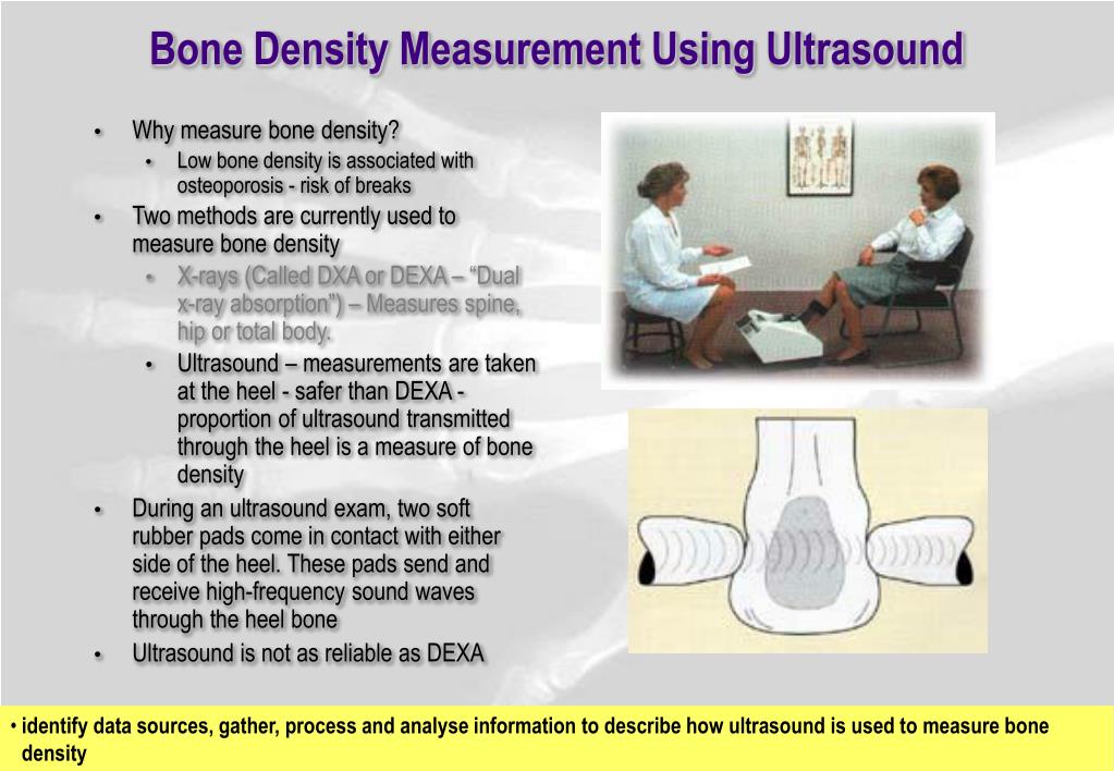
Describe How Ultrasound Is Used to Measure Bone Density
SOS is influenced by the elasticity of bone as well as by its density. A portable bone density scan is not as precise because only one bone site is tested.
Ppt Medical Physics Ultrasound Powerpoint Presentation Free Download Id 5588587
Learn more about bone density exams in our Health Library.
. Doctors use DXA scans most widely. In the central DXA examination which measures bone density of the hip and spine the patient lies on a padded table. The test measures the quantity of bone at specific locations in the body namely the mid-spine bones lumbar region and the upper end of the thighbone where it connects to the pelvis total hip and its.
Absorption predominates in cortical bone and scattering in trabecular bone. The test uses X-rays to measure how many grams of calcium and other bone minerals are packed into a segment of bone. The way we live shapes our bones.
Ultrasound imaging could help boost osteoporosis screening. These tests are often used for screening purposes and can help identify people who might benefit from follow-up bone density testing at the hip and lumbar spine. There are some portable DEXA or ultrasound units used for general screenings that examine the heel lower arm or wrist.
The most widely used is a scan called dual energy X-ray absorptiometry DXA or. The most common variable reflecting ultrasound attenuation through bone is known as broadband ultrasound attenuation BUA that is a measure of the frequency dependence of the attenuation of the. There are a lot of things we can do to help keep our bones healthy.
BUA is a measure of the approximately linear frequency dependence of ultrasound attenuation. Bone mineral density BMD gauges. The ultrasound wave is produced in the form of a sinusoid impulse by special piezoelectric probes and is detected once it has passed through the medium.
About heel ultrasound machines. The machine works by measuring how sound waves move through the bone in the heel. However as previous studies showed that the BUA is sensitive to repositioning and soft tissue the reproducibility of measurement will be difficult to achieve.
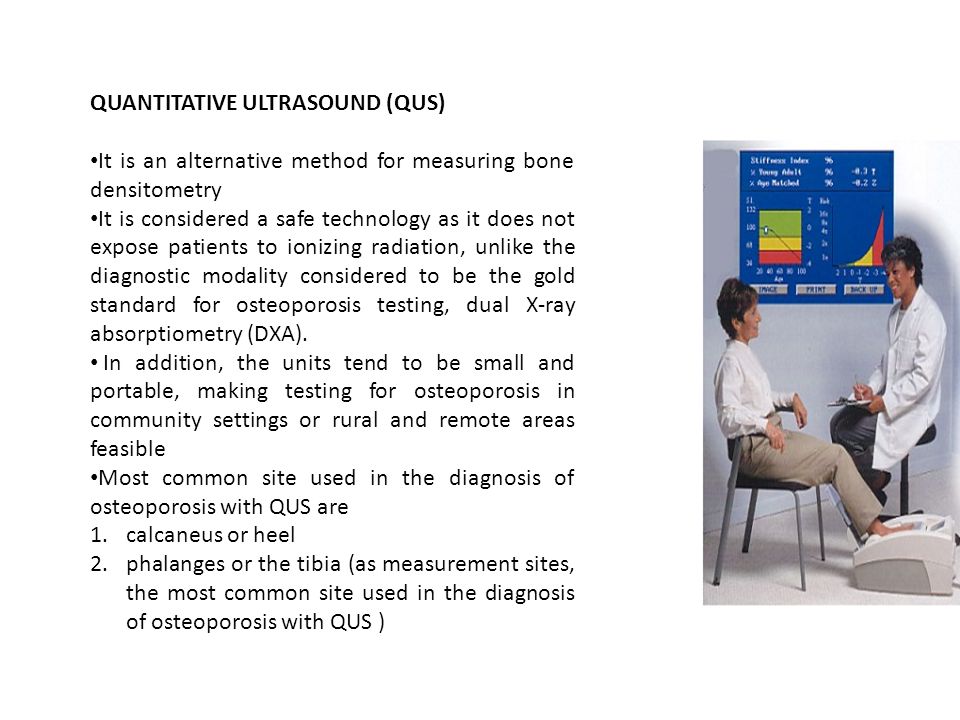
Bone Densitometry Principles Current techniques use ionizing radiation or ultrasound Most common is dual-energy x-ray absorptiometry DXA DXA Advantages Low radiation dose Readily available Simple to use Short scan time High-resolution images Precise Stable calibration DXA Subtraction Technique. For various reasons the DEXA scan is considered the gold standard or most accurate test. There are limited guidelines for interpreting ultrasound tests to predict fracture risk or diagnose osteoporosis.
X-ray scans CT scans and ultrasound scans can measure a persons bone density. What does the test do. In our system we used Net Delay Time NDT value pressure detection and.
Weak x-rays are usually sent from below through the bones of the spine and the top of the thigh bone. BUA is determined by mechanisms of diffraction scattering and absorption in the bone marrow and soft tissue. September 28 2019.
This is the method used to determine efficacy in the recent large clinical trials and to characterize fracture risk in large epidemiological studies. A DXA scan is preferred over ultrasound for measuring bone density. A bone density test is used to measure bone mineral content and density.
Bone density is commonly measured using a special x-ray technique called dual energy x-ray absorptiometry DXA or DEXA. These results are combined to give the Quantitative Ultrasound Index QUI or Stiffness. The QUS method involves generating ultrasound impulses that are transmitted transversally or longitudinally through the bone being studied.
The thought is that sound waves travel. A bone density test determines if you have osteoporosis a disorder characterized by bones that are more fragile and more likely to break. Bone density tests provide a precise measure of whether you have osteopenia or osteoporosis.
Recently much attention has been paid to the use of ultrasound to detect bone density as it is non-ionizing relatively inexpensive and simple to use. To assess the hip the patients foot is placed in a. This technique can be used to measure bone density in the spine hip forearm and the total body.
When bones are somewhat thin the condition is called osteopenia. Bone mineral density BMD based on an ultrasound measurement of the calcaneus. For these reasons ultrasound is normally only used as a bone density test when DXA scans are not available.
It may be done using X-rays dual-energy X-ray absorptiometry DEXA or DXA or a special CT scan that uses computer software to determine bone density of the hip or spine. In the last years quantitative ultrasound QUS methods have been developed to assess bone mineral status in some peripheral skeletal sites such as calcaneus phalanges of the hand and tibia. A bone density testalso referred to as a DXA scan or a bone mineral density BMD testis the current way to diagnose osteoporosis.
It is one of the most common methods to determine bone density as it is fast and highly accurate. An x-ray generator is located below the patient and an imaging device or detector is positioned above. If someone has a lot of wear and tear in their spine this approach is only used to look at their thigh bone.
The broadband ultrasound attenuation BUA and the speed of sound SOS are usually measured to evaluate the bone density. It does not actually measure bone mineral density but speed of sound SOS in metressecond and broadband ultrasound attenuation BUA in decibelsmegahertz. Peripheral bone density tests measure bone density in the lower arm wrist finger or heel.
The bones that are most commonly tested are in the spine hip and sometimes the forearm. Request an appointment phone 443-997-7237. There are benefits and limitations to each of the different bone density tests.
Testing your bone density -- how strong your bones are -- is the only way to know for sure if you have osteoporosis. When bones become very thin the condition is called osteoporosis. Quantitative ultrasound which does not involve x-rays may also be used.
Usually the detection is. In this cross-sectional study the bone mineral density of the calcaneus was investigated in healthy young n 35 22-33 years and middle-aged n 49 45-59 years men. Ultrasound machines became popular as a screening tool for osteoporosis because they are portable not too expensive involve no radiation and do not require a licensed technician to operate.
To assess the spine the patients legs are supported on a padded box to flatten the pelvis and lower spine. Dual energy x-ray absorptiometry DEXA x-ray beams of differing energy are used to detect bone and soft tissue density separately. Ultrasound Techniques DEXA Several methods are available to measure bone density but currently the most widely used technique is DEXA Dual Energy Xray Absorptiometry.
QUS techniques are safe easy to use radiation-free and devices are portable so that they are particularly indicated to assess bone mineral status in children. A BMD test measures your bone mineral density and compares it to that.

Quantitative Ultrasound Method A Ultrasound Beam Through A Bone Download Scientific Diagram

Quantitative Ultrasound Method A Ultrasound Beam Through A Bone Download Scientific Diagram
Quantitative Ultrasound Qus What Is Ultrasound Sound Waves Of Extremely High Frequency Inaudible To The Human Ear Ultrasound Can Be Used To Examine Ppt Download
No comments for "Describe How Ultrasound Is Used to Measure Bone Density"
Post a Comment